Meniscectomy is a surgical procedure for patients with a torn meniscus where conservative treatments have failed to relieve pain and mechanical symptoms. Meniscectomy is recommended based on the ability of meniscus to heal, patient’s age, health status, and activity level.
The meniscus is the C-shaped two pieces of cartilage located between thighbone and shin bone that act as shock absorbers and cushion the joints. Meniscus distributes the body weight uniformly across the joint and avoids the pressure on any one part of the joint and development of arthritis. Being the weight-bearing part, the meniscus is prone to wear and tear and meniscal tear is one of the common knee injuries. Meniscal tear may be developed by people of all ages and is more common in individuals who play contact sports.
On the pattern of tear, meniscal tear may be of different types such as longitudinal, parrot-beak, flap, bucket handle, and mixed/complex tear. Sudden twisting, squatting, or being tackled may cause a meniscal tear in adults and natural ageing and degeneration of the meniscal tissue may cause the tear in elderly individuals. Meniscal tear may cause severe pain, stiffness, and swelling, catching or locking of the knee, and may limit the movement. Meniscal tear is often diagnosed with the presenting symptoms and imaging techniques such as magnetic resonance imaging scan.
Treatment
Conservative treatments for meniscal tear include R.I.C.E (Rest, Ice, Compression, and Elevation) and use of non-steroidal anti-inflammatory medications. Surgery is recommended in severe cases and will be performed using arthroscopic (keyhole) technique. Your surgeon may also order a rehabilitation program following the surgery.
What happens in an Arthroscopic Meniscectomy?
- The arthroscope is a small fibre-optic viewing instrument made up of a tiny lens, light source, and video camera. The surgical instruments used in arthroscopic surgery are very small (only 3 or 4 mm in diameter), but appear much larger when viewed through an arthroscope.
- The camera attached to the arthroscope displays the image of the joint on a television screen, allowing the surgeon to look throughout the knee at cartilage and ligaments, and under the kneecap.
- Then the surgeon makes two small incisions (about 1/4 of an inch), around the knee joint area. Each incision is called a portal. In one portal, the arthroscope is inserted to view the knee joint.
- Along with the arthroscope, a sterile solution is pumped into the joint which expands the viewing area, giving the surgeon a clear view and room to work. The other portal is used for the insertion of tiny surgical instruments.
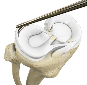
- With the images from the arthroscope as a guide, your surgeon can look at the menisci and confirm the type, location, and extent of the tear. Once your surgeon has located the meniscal tear, surgical scissors and shavers are inserted into the portals to remove the torn menisci.
- In total meniscectomy, entire menisci are removed and in partial meniscectomy, only the torn part of the tissue is removed leaving the intact tissue in place with edges smoothened.

